Spondylosis refers to degenerative arthritis of the spine including osteoarthritis of the discovertebral and facet joints and degenerative changes of related soft tissues including surrounding ligaments and muscles.
The cause of spondylosis remains unclear. A widely cited hypothesis states that degenerative changes begin with the loss of water content in the annulus fibrosis which then becomes drier and weaker and eventually, the disc content leaks out resulting in intervertebral disc potrusion and narrowing of the disc space. this subsequently leads to increased mechanical stress at the discovertebral joints, the facet joints and the spinal ligaments causing both bony overgrowth and ligament thickening (see Fig 1). Ref [1].

Spondylolysis refers to a bone defect in the pars interarticularis (‘bridge between the joints’) which acts as an isthmus or bony bridge between the inferior and superior articular surfaces of the neural arch of a single vertebra. When bilateral spondylolysis is present, the posterior aspect of the neural arch, including the inferior articular surfaces, is no longer connected by bone to the rest of vertebra.
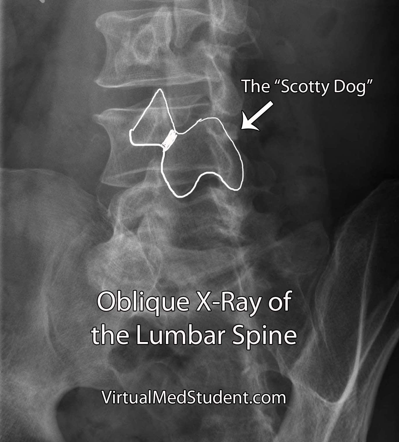
Spondylolysis is an acquired condition. It mostly occurs at the L5 vertebra (where about 90% of the cases are found). It is more common in males and females with roughly a 2:1 ratio. The most likely etiology is a stress fracture of the pars that persists as a nonunion. Lateral oblique radiographs may reveal the classic ‘collar on the Scottie dog’ finding (see Fig 2).
Bilateral occurences are more common than unilateral presentations and is usually more prevalent among adolescents and young adults who aggressively participate in sports requiring repetitive flexion-extension movements such as gymnastics, throwing sports, football etc.
Spondylolisthesis refers to displacement of a vertebral body in relation to the one below it. The most common type of spondylolisthesis and the alignment abnormality that is implied when the term is used is an anterior displacement AKA anterolisthesis (Fig 3). However, spondylolisthesis can also occur in the posterior direction (retrolisthesis) as well as in a lateral direction (laterolisthesis).
Spondylolisthesis is classified by etiology (dysplastic, isthmic, degenerative, traumatic or pathologic) and by grade (grade I – IV). Spondylolisthesis is an abnormal finding and whenever present, is an indication that there is structural and functional failure of the neural arch and facet joints which are responsible for maintaining normal vertebral alignment. Ref [2].

[1] Li S. and Yao J. Spinal Imaging and Image Analysis: Lecture Notes in Computational Vision and Biomechanics 18. 2014. Springer Publishers. Part I. Clinical Imaging and Applications. Arthritis of the Spine. Pg 31.
[2] Frontera WR et al. Essentials of Physical Medicine and Rehabilitation. Musculosketal Disorders, Pain and Rehabilitation. 2015. 3rd Edition. Elsevier Saunders Publishers. Chapter 49. Lumbar Spondylolysis and Spondylolisthesis. Pg 249.
0
0